.png?width=80&height=80&name=Clove%20Dental%20White%20Small%20(28).png "Clove Dental White Small (28)")
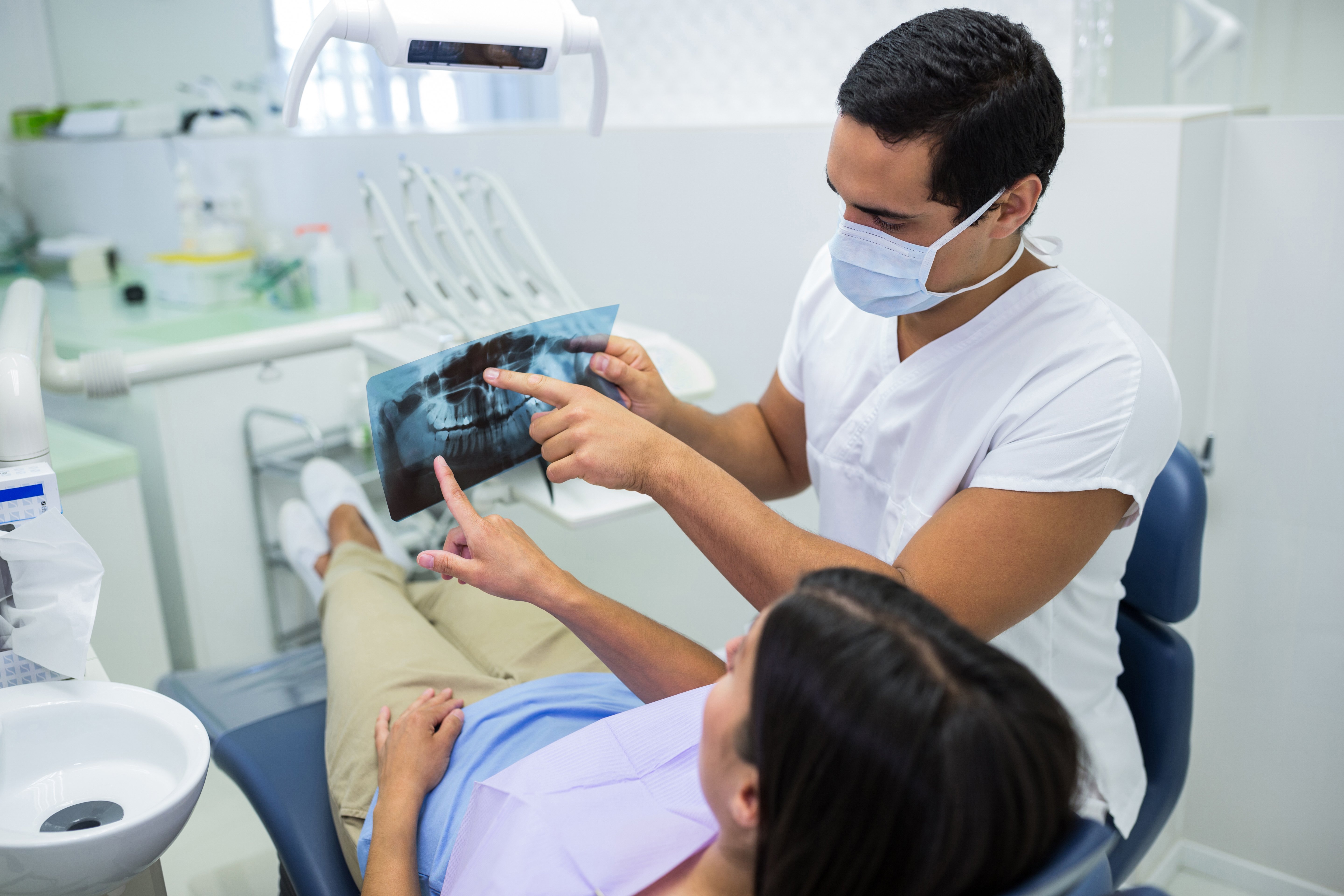
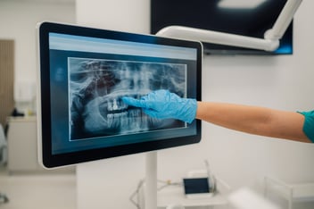
Dental X-rays are considered safe when taken for appropriate clinical reasons at recommended intervals. A dentist in Camarillo using modern digital X-ray equipment exposes patients to significantly less radiation than traditional film X-rays, the equivalent of a few hours of natural background radiation or less than a two-hour flight. The radiation from a full-mouth series of digital dental X-rays is a fraction of the daily environmental radiation most people receive from natural sources.
Key Takeaways
- Modern digital dental X-rays expose patients to less radiation than traditional film. The exposure from routine dental imaging is comparable to a few hours of everyday background radiation.
- The primary protective equipment, lead apron and thyroid collar is routine practice and further reduces any incidental exposure.
- The risk of not taking clinically indicated X-rays, missing early decay, bone loss or pathology, outweighs the minimal radiation exposure associated with modern dental imaging.
Most patients who decline or question dental X-rays are making a reasonable health-protective calculation, they're concerned about radiation exposure and wondering whether the images are really necessary this time. It's a legitimate question, and a dentist in Camarillo who takes it seriously rather than dismissing it provides a better standard of care than one who simply insists.
The answer to "are dental X-rays safe?" is yes, with two important qualifications: the X-rays should be taken with modern equipment and they should be taken for clinical reasons that justify them, not as routine regardless of patient history. At Clove Dental, we approach X-ray decisions exactly this way.

"Didn't I Already Have an X-Ray?" The Question Patients Ask Most Often
This question comes from patients who feel like they're being imaged every visit and sometimes they're right to ask whether the frequency is warranted.
The answer depends on what the previous X-rays showed, how much has likely changed since, and what clinical questions remain unanswered without new imaging. A patient with a history of frequent cavities and several new fillings in the last two years needs more regular X-ray monitoring than a patient who has had clean checkups for a decade. A patient being seen for the first time at a new practice needs baseline imaging to give the new dental team the clinical picture they don't yet have.
X-rays aren't ordered on a fixed calendar regardless of clinical context. They're ordered when the information they provide is expected to change or inform treatment decisions which means frequency is calibrated to the individual patient.
What Your Dentist Is Actually Looking for That Can't Be Seen During an Exam
The visual examination that happens during a dental checkup is limited to what's visible above the gum line and accessible by direct observation. It can identify visible staining, surface wear, obvious chips and gum tissue changes. It cannot show:
- Cavities developing between teeth, the most common location for new decay in adults.
- Bone levels around tooth roots, the primary measure of periodontal health.
- Conditions developing inside or beneath existing restorations.
- Cysts, abscesses or pathology within the bone.
- Root tip infections that haven't yet created visible swelling or symptoms.
Each of these can be present, progressing, and causing cumulative damage while producing no symptoms and nothing visible to clinical examination. X-rays are the diagnostic tool that extends clinical observation into these invisible spaces, not an add-on but a fundamental component of a complete dental examination.
Why Modern Dental X-Rays Are Different From What Many People Remember
The dental X-rays most patients over 40 remember from childhood involved film that required chemical processing, took longer to capture, and used more radiation to achieve adequate image quality than current technology requires.
Digital X-ray sensors, now standard in most dental offices, require dramatically less radiation to produce an image than traditional film. The exposure from a full set of digital bitewing X-rays is typically less than 0.005 millisieverts, a fraction of the average 3 millisieverts of background radiation a person in the United States receives annually from natural sources (radon, cosmic radiation, soil minerals).
To put it in more tangible terms: the radiation from a complete set of digital dental X-rays is comparable to approximately 1 to 2 hours of background radiation during normal daily activity or the additional radiation exposure of a short domestic flight. The comparison to other medical imaging is equally reassuring, a dental full-mouth series delivers less radiation than a single chest X-ray.
The Safest X-Ray Is the One That's Taken for the Right Reason
This is the most important principle in dental X-ray safety: the decision to take an X-ray should be a clinical one, made because the information expected from the image is valuable for the patient's care.
An X-ray taken because the schedule says so, without a clinical indication that the information will change the assessment or treatment plan, is unnecessary regardless of how low the radiation dose is. The goal is to find problems early, not to demonstrate thoroughness.
At Clove Dental, X-ray recommendations follow this standard: the clinical need drives the decision, and patients who want to understand why a specific image is being recommended will always receive a clear explanation.
Conclusion
Dental X-rays are among the safest diagnostic imaging procedures in medicine, particularly with modern digital equipment. The radiation exposure is minimal, the protective measures are routine, and the clinical value of the information they provide, particularly for conditions that develop silently, is significant.
At Clove Dental, our dentist in Camarillo team takes X-rays when they're clinically indicated and explains why.
FAQs
Do I have the right to decline dental X-rays?
Yes, patients can decline X-rays. A dentist in Camarillo should respect that decision while ensuring the patient understands what clinical information will be unavailable as a result. Some conditions are only detectable with imaging; a patient who declines X-rays may have limited diagnostic information available at that visit.
Are dental X-rays safe during pregnancy?
With appropriate shielding (lead apron and thyroid collar), dental X-rays are considered safe during pregnancy for clinically necessary imaging. Elective or non-urgent X-ray series are typically deferred until after delivery.
Why does my dentist use a thyroid collar?
The thyroid gland is sensitive to radiation and sits near the area imaged during dental X-rays. A thyroid collar, a wrap of lead shielding around the neck, reduces any incidental scatter radiation exposure to this organ during imaging.
How often should I have dental X-rays taken?
The appropriate frequency depends on your cavity risk, gum disease history, and what was found at prior visits. Most low-risk adults receive bitewing X-rays every 18 to 36 months. Higher-risk patients may need annual bitewings.
Are digital X-rays really safer than traditional film?
Yes, digital X-ray sensors require approximately 50 to 80 percent less radiation than traditional film to produce a comparable image. They also produce immediate results without chemical processing and can be digitally enhanced to improve diagnostic clarity.